At most medical schools, first-year anatomy is about diagrams, labels, and memorization. But at Touro College of Osteopathic Medicine (TouroCOM), anatomy becomes alive. From day one, students are challenged to apply what they learn in class to real-life clinical scenarios—making anatomy feel less like a lecture, and more like a diagnosis waiting to happen.
Cadaver Lab: The First Clinical Teacher
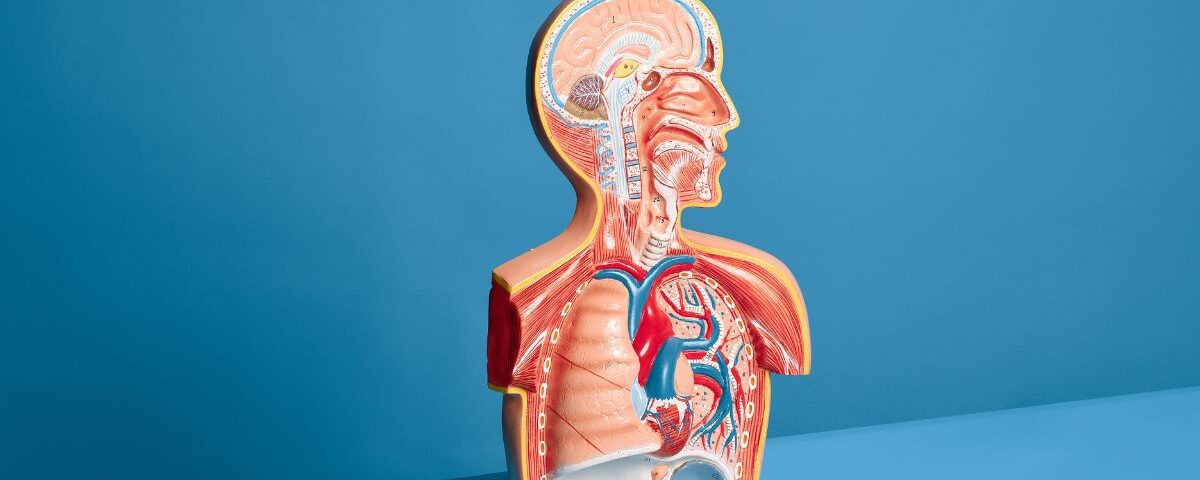
The cadaver lab isn’t just an academic rite of passage at TouroCOM—it’s the first chance to encounter the human body as future physicians. Every muscle, nerve, and organ becomes a clue to how the body functions in health and breaks down in disease.
Students are encouraged to ask:
- What would happen if this artery were blocked?
- Could this nerve injury explain foot drop?
- Is this what scoliosis would look like in real life?
It’s not about passing a test—it’s about clinical thinking from the very beginning.
Case-Based Learning in First Year
TouroCOM integrates case-based learning early on. After covering the brachial plexus in anatomy, students might tackle a clinical case like:
“A 32-year-old motorcycle crash victim presents with arm weakness and numbness. What’s injured?”
Suddenly, those complicated nerve roots are no longer abstract—they’re the key to a diagnosis. And when students understand why the patient can’t lift their arm or where the numbness stops, they start thinking like doctors.
Anatomy Meets Radiology: Learning to Read Images Early
First-years at TouroCOM are introduced to radiologic anatomy—learning to read X-rays, CT scans, and MRIs alongside gross anatomy. This dual approach allows students to recognize:
- Fractures and dislocations
- Organ enlargement
- Brain bleeds and midline shifts
By the time they reach rotations, they’re confident and competent in identifying key findings that would stump many first-year residents.
Real Stories, Real Students, Real Learning
Sarah, a current OMS-I student at TouroCOM, recalls her first anatomy-related “aha!” moment:
“We were learning about the neck muscles, and I read about a patient with torticollis. Suddenly, I wasn’t just memorizing the sternocleidomastoid—I was seeing how it could cause a child’s head tilt. It made me want to learn more.”
These moments are common at TouroCOM, where theory meets practice in every lecture, lab, and case review.
OMM and Anatomy: A Natural Pair
As a school of osteopathic medicine, TouroCOM emphasizes Osteopathic Manipulative Medicine (OMM)—and that means students must know the body deeply.
- Palpation skills rely on muscle origin and insertion points
- Joint mobilization depends on understanding connective tissue anatomy
- Visceral manipulation builds on organ position and structure
Anatomy in action isn’t just in diagnosis—it’s in treatment too.
Why It Matters?
Medical knowledge doubles every few years, but the human body doesn’t change. Anatomy is the timeless foundation of medicine, and at TouroCOM, it’s taught in a way that prepares students to:
- Think clinically, not just academically
- Work confidently in surgical, primary care, and emergency settings
- Communicate clearly with patients and colleagues about bodily systems
Conclusion
TouroCOM’s anatomy curriculum isn’t about endless memorization—it’s about activation. By integrating real cases, radiology, and OMM into every stage of learning, students transform from passive learners into proactive clinicians.
Because in medicine, knowing what is where matters. But knowing why it hurts—and how to fix it—starts with anatomy in action.
FAQs
Q: Do students start clinical cases in their first year at TouroCOM?
Yes! Case-based learning begins early, especially alongside anatomy topics.
Q: How does anatomy help with OMM?
Understanding precise structure is essential for safe and effective hands-on treatment.
Q: Is radiologic anatomy part of the first-year curriculum?
Absolutely. Students learn to interpret basic scans and apply that to clinical thinking.