

For many medical students, neuroanatomy feels like one of the most intimidating courses. The brain’s intricate structures, endless tracts, and weird Latin names can make your head spin. But here’s the twist: once you understand how the brain is wired, you can start diagnosing smarter and faster.
This isn’t just about passing exams. Knowing your brain maps—from the motor cortex to cranial nerves—translates into real clinical power. It helps you pinpoint where a lesion might be, what symptoms to expect, and even what treatments to consider.
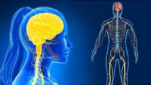
Brain Maps: The GPS of Clinical Diagnosis

Think of brain maps as the brain’s internal GPS system. They show how functions like movement, sensation, and speech are organized. When something goes wrong, these maps tell you where to look.
Example Table: Common Brain Regions and Clinical Correlations
| Brain Region | Function | Clinical Sign When Damaged |
|---|---|---|
| Broca’s Area | Speech production | Expressive aphasia |
| Primary Motor Cortex | Voluntary movement | Contralateral paralysis |
| Cerebellum | Coordination & balance | Ataxia, intention tremor |
| Occipital Lobe | Vision processing | Visual field defects |
| Wernicke’s Area | Language comprehension | Receptive aphasia |
By studying these areas, you develop a functional framework. You’re no longer just memorizing anatomy—you’re learning how to track symptoms back to brain structures.
Real-Life Case: When Neuroanatomy Saves the Day
Let’s say a patient comes in with:
- Slurred speech
- Right facial droop
- Weakness in the right arm and leg

If you know your neuroanatomy, your brain will automatically think: “Left internal capsule or motor cortex involvement.” You can now confidently suggest imaging and even start discussing differentials like a stroke in the MCA territory.
Without this anatomical insight, you’d be lost in guesswork.
Smarter Diagnoses Start with Smarter Studying
If you’re still in the memorization stage, here are some quick ways to make your neuroanatomy stick in a real-world way:
1. Use Clinical Cases While Studying
Match each anatomical structure with a real clinical symptom.
2. Draw the Maps Yourself
Color-coded motor/sensory maps (like the homunculus) boost retention and understanding.
3. Practice Localizing Lesions
Challenge yourself with questions like, “Where in the brain could this lesion be if the patient presents with X, Y, and Z?”
From Textbook to Touchpoint: Why Neuroanatomy is Your Clinical Superpower
Here’s the truth: great clinicians aren’t just good at pattern recognition—they understand why those patterns exist. Neuroanatomy bridges the gap between symptom and source.
Every neurologic exam, every MRI, and every confused patient becomes clearer when you can “see” the underlying brain map in your mind.
Conclusion:
Understanding neuroanatomy isn’t just about acing exams or surviving lab practicals. It’s about becoming the kind of clinician who can see what others miss.
When neuroanatomy meets real life, something magical happens: you stop guessing and start diagnosing smarter. You’re not just learning where things are—you’re learning how they work together in the real human body.
FAQs
Q1: Do all medical specialties require deep knowledge of neuroanatomy?
A: No, but specialties like neurology, neurosurgery, and psychiatry rely on it heavily. Even general practitioners benefit from basic neuroanatomy skills.
Q2: What’s the best way to learn brain maps effectively?
A: Active recall using clinical scenarios and diagrams is one of the most effective methods.
Q3: How early in med school should I start connecting neuroanatomy to clinical practice?
A: As early as possible. Even basic understanding helps you think critically during rotations and exams.